Results ready 4-6 hours (after completion of the test).
Extra
SCAN TIME: 30 MINUTES
Setup
All working days
Test in Brief
An imaging test that uses powerful magnets and radiowaves to produce images. Unlike X-rays and CT scan, it does NOT use ionizing radiation. The patient lies on a motorized table which will move the patient into the scanner which resembles a giant doughnut (like a tunnel that is open at both ends).
Exam Requirements
Remove all metal objects (e.g. watches, jewellery); patient should complete the safety questionnaire that is provided prior to the procedure; the scanner is a tunnel that is open at both ends; headphones or earphones will be used to muffle the sounds produced by the scanner. Occasionally, a special dye (contrast media) is administered through the vein. Patient should lie still till the end of procedure/exam to avoid getting blurry images.
Instructions For Referring Doctor Or Institution
Please state clinical indication for the test on the request form; for NHIF & other insurance clients, provide the necessary documentation in full inclusive of stamps and referrer’s name/ signature/ board registration details where required. In case of NHIF queries/ clarifications, call 0785 033335 (Nakuru).
Instructions For Radiographer
Guide the patient when filling the safety questionnaire to identify any contra-indications before the procedure/exam.
Method
Patient is placed in the MRI magnetic bore; radiofrequency is sent in depending on the sequence/ time; radiofrequecy is turned off; images are reconstructed on the MRI Console & assessed for good radiographic quality. Images are then reviewed by the radiologist. A comprehensive report detailing the patient name, gender, age, procedure date, procedure name and findings is provided.
Usefulness /Advantages
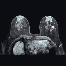
MRI, used with mammography and breast ultrasound, can be a useful diagnostic tool. Recent research has found that MRI can locate some small breast lesions sometimes missed by mammography. It can also help detect breast cancer in women with breast implants and in younger women who tend to have dense breast tissue.