Report
Results ready 4-6 hours (after completion of the test).
Extra
SCAN TIME: 30 MINUTES
Setup
All working days
Test in Brief
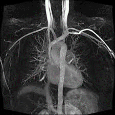
Complete patient history with relevant lab investigations including recent UECs and concurrent Egfr results/documents.
Exam Requirements
Remove all metal objects (e.g. watches, jewellery); patient should complete the safety questionnaire that is provided prior to the procedure; the scanner is a tunnel that is open at both ends; headphones or earphones will be used to muffle the sounds produced by the scanner. Occasionally, a special dye (contrast media) is administered through the vein. Patient should lie still till the end of procedure/exam to avoid getting blurry images.
Instructions For Referring Doctor Or Institution
Please state clinical indication for the test on the request form; for NHIF & other insurance clients, provide the necessary documentation in full inclusive of stamps and referrer’s name/ signature/ board registration details where required. In case of NHIF queries/ clarifications, call 0785 033335 (Nakuru).
Instructions For Radiographer
Guide the patient when filling the safety questionnaire to identify any contra-indications before the procedure/exam.
Method
Patient is placed in the MRI magnetic bore; radiofrequency is sent in depending on the sequence/ time; radiofrequecy is turned off; images are reconstructed on the MRI Console & assessed for good radiographic quality. Images are then reviewed by the radiologist. A comprehensive report detailing the patient name, gender, age, procedure date, procedure name and findings is provided.
Usefulness /Advantages
Adding contrast makes it possible for the radiologist to detect even the smallest tumor and provides information about the precise location of the tumor. The radiologist can interpret an MRI contrast scan better, since they have more clarity and generate better-quality images.