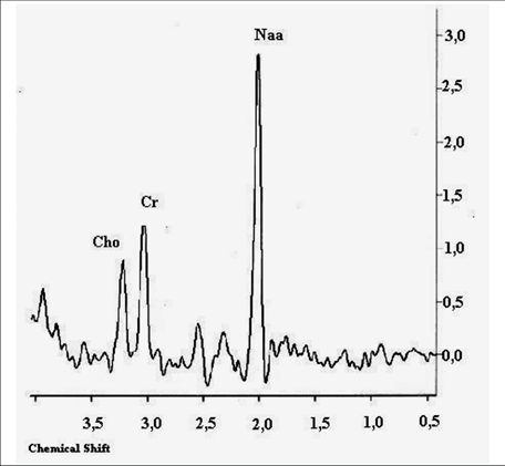
MRS(SPECTROSCOPY)
MRI spectroscopy is known as magnetic resonance spectroscopy (MRS). It is used to evaluate the presence and concentration of various metabolites in tumours. MRS has been applied as both a research and a clinical tool in order to detect visible or nonvisible abnormalities. The adaptability of MRS allows a technique that can probe a wide variety metabolic uses across different tissues. Although MRS is mostly applied for brain tissue, it can be used for detection, localization, staging, tumor aggressiveness evaluation, and tumor response assessment of breast, prostate, hepatic, and other cancers.
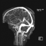
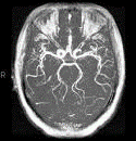
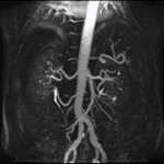
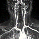
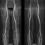
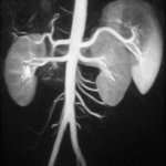
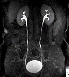
MRI ANGIOGRAPHY
Magnetic resonance angiography is a group of techniques based on magnetic resonance imaging to image blood vessels. Magnetic resonance angiography is used to generate images of arteries and veins in order to evaluate them for stenosis, occlusions, aneurysms, thrombosis or other abnormalities. It is useful in evaluating the blood vessels in various systems; cerebral, carotids, upper limbs, chest, abdominal, pelvic, cardiac and lower limbs.
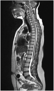
MRI SPINE
MRI spine is useful in evaluating symptoms such as neck pain, upper and lower back pain, numbness, tingling or weakness in the lower limbs, arms, shoulders, or neck area.
In addition, it is useful in evaluating congenital birth defects, spine deformities as well as pre and post surgical follow up.
Tumors, bleeding, swelling, infections, or inflammatory conditions in the vertebrae or surrounding tissues can also be evaluated using MRI.
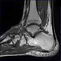
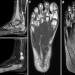
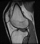
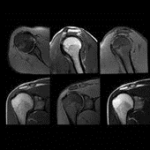
MRI EXTREMITIIES
An extremity MRI is a type of scan used specifically for diagnostic imaging of the arm, leg, hand, or foot. The machine uses radio waves and a magnetic field to generate images of the inside of the extremity in order to diagnose problems with the muscles, bones, joints, nerves, or blood vessels.
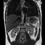
MRCP (MR Cholangiopancreatography)
Magnetic resonance cholangiopancreatography or MRCP uses a powerful magnetic field, radio waves and a computer to evaluate the liver, gallbladder, bile ducts, pancreas and pancreatic duct for disease. It is noninvasive and does not use ionizing radiation

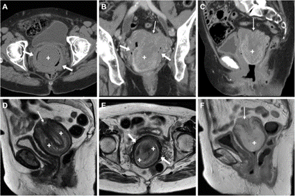
MRI FOR BOTH GYNAECOLOGICAL AND UROLOGICAL CONDITIONS
MRI does not work very well in the urinary tract. Its signals will not show calcifications in soft tissue and bladder abnormalities.
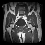
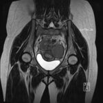
MRI FOR GYNAECOLOGICAL CONDITIONS
In a female with pelvic pain, MRI can accurately identify adenomyosis, enumerate and localize uterine fibroids, and provide more accurate identification of endometriosis and masses of the ovary and overall condition of the female reproductive system.
MRI MYELOGRAM
It a is type of MRI examination that uses a contrast medium and magnetic resonance imaging scanner to detect pathology of the spinal cord, including the location of a spinal cord injury, cysts, tumors and other abnormalities.
PAEDIATRIC MRI STUDIES
Children’s magnetic resonance imaging (MRI) uses a powerful magnetic field, radio waves and a computer to produce detailed pictures of the inside of your child’s body. MRI may be used to help diagnose or monitor treatment for a variety of conditions within the brain, chest, abdomen, pelvis and extremities.
Images are clear, especially for soft tissue, less invasive, and timely.
No radiation is an added advantage for pediatrics, hence recommend MRI more often.