MRI RENAL ANGIOGRAM

Category

Routine Imaging

Synonyms/Aliases

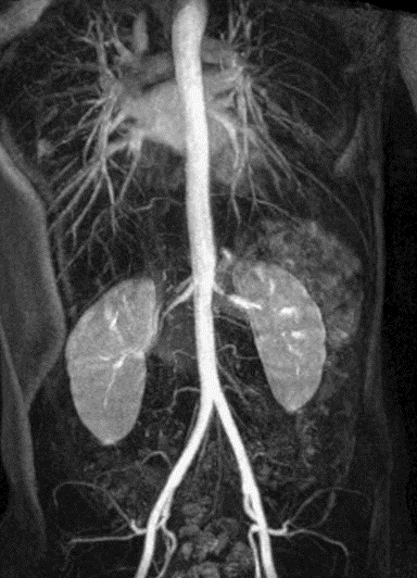
MRI OF THE KIDNEYS, MRI RENAL ARTERIOGRAM

Abbreviations

- N/A

Report

4-6 hours (after completion of the test).

Setup

All working days

Scan

45 MINUTES

Test in Brief

Capture accurate patient information when filling the questionnaire to identify indications and contra-indications before the procedure/exam.

Exam Requirements

Full bladder prior to procedure.Remove all metal objects (e.g. watches, jewellery); patient should complete the safety questionnaire that is provided prior to the procedure; the scanner is a tunnel that is open at both ends; headphones or earphones will be used to muffle the sounds produced by the scanner. Occasionally, a special dye (contrast media) is administered through the vein. Patient should lie still till the end of procedure/exam to avoid getting blurry images.

Instructions

INSTRUCTIONS FOR REFERRING DOCTOR OR INSTITUTION: Please state clinical indication for the test on the request form; for NHIF & other insurance clients, provide the necessary documentation in full inclusive of stamps and referrer’s name/ signature/ board registration details where required. In case of NHIF queries/ clarifications, call 0785 033335 (Nakuru). INSTRUCTIONS FOR RADIOGRAPHER: Guide the patient when filling the safety questionnaire to identify any contra-indications before the procedure/exam.

Method

Patient is placed in the MRI magnetic bore; radiofrequency is sent in depending on the sequence/ time; radiofrequecy is turned off; images are reconstructed on the MRI Console & assessed for good radiographic quality. Images are then reviewed by the radiologist. A comprehensive report detailing the patient name, gender, age, procedure date, procedure name and findings is provided.

Usefulness /Advantages

MRI scans of your kidneys can help see conditions like kidney cancer, chronic kidney disease, renal vein thrombosis, and the presence of tumors, masses, stones, or infection. There are two types of MRI scans for your kidneys: imaging with or without contrast agents.

MRI PROSTATE

Category

Routine Imaging

Synonyms/Aliases

MRI PELVIS

Abbreviations

- N/A

Report

4-6 hours (after completion of the test).

Setup

All working days

Scan

30 MINUTES

Test in Brief

Capture accurate patient information when filling the questionnaire to identify indications and contra-indications before the procedure/exam.

Exam Requirements

Full bladder prior to procedure.Remove all metal objects (e.g. watches, jewellery); patient should complete the safety questionnaire that is provided prior to the procedure; the scanner is a tunnel that is open at both ends; headphones or earphones will be used to muffle the sounds produced by the scanner. Occasionally, a special dye (contrast media) is administered through the vein. Patient should lie still till the end of procedure/exam to avoid getting blurry images.

Instructions

INSTRUCTIONS FOR REFERRING DOCTOR OR INSTITUTION: Please state clinical indication for the test on the request form; for NHIF & other insurance clients, provide the necessary documentation in full inclusive of stamps and referrer’s name/ signature/ board registration details where required. In case of NHIF queries/ clarifications, call 0785 033335 (Nakuru). INSTRUCTIONS FOR RADIOGRAPHER: Guide the patient when filling the safety questionnaire to identify any contra-indications before the procedure/exam.

Method

Patient is placed in the MRI magnetic bore; radiofrequency is sent in depending on the sequence/ time; radiofrequecy is turned off; images are reconstructed on the MRI Console & assessed for good radiographic quality. Images are then reviewed by the radiologist. A comprehensive report detailing the patient name, gender, age, procedure date, procedure name and findings is provided.

Usefulness /Advantages

to evaluate prostate cancer and see if it is limited to the prostate. MRI provides information on how water molecules and blood flow through the prostate. This helps determine whether cancer is present and, if so, whether it is aggressive and if it has spread.

MRI PERIPHERAL ANGIOGRAM

Category

Routine Imaging

Synonyms/Aliases

MRI LOWER/ UPPER EXTREMITIES ANGIOGRAMS

Abbreviations

MRI P.A

Report

4-6 hours (after completion of the test).

Setup

All working days

Scan

45 MINUTES

Test in Brief

Capture accurate patient information when filling the questionnaire to identify indications and contra-indications before the procedure/exam.

Exam Requirements

Remove all metal objects (e.g. watches, jewellery); patient should complete the safety questionnaire that is provided prior to the procedure; the scanner is a tunnel that is open at both ends; headphones or earphones will be used to muffle the sounds produced by the scanner. Occasionally, a special dye (contrast media) is administered through the vein. Patient should lie still till the end of procedure/exam to avoid getting blurry images.

Instructions

INSTRUCTIONS FOR REFERRING DOCTOR OR INSTITUTION: Please state clinical indication for the test on the request form; for NHIF & other insurance clients, provide the necessary documentation in full inclusive of stamps and referrer’s name/ signature/ board registration details where required. In case of NHIF queries/ clarifications, call 0785 033335 (Nakuru). INSTRUCTIONS FOR RADIOGRAPHER: Guide the patient when filling the safety questionnaire to identify any contra-indications before the procedure/exam.

Method

Patient is placed in the MRI magnetic bore; radiofrequency is sent in depending on the sequence/ time; radiofrequecy is turned off; images are reconstructed on the MRI Console & assessed for good radiographic quality. Images are then reviewed by the radiologist. A comprehensive report detailing the patient name, gender, age, procedure date, procedure name and findings is provided.

Usefulness /Advantages

an excellent method for diagnosing the anatomic location and degree of stenosis in PAD, stenosis ,aneurysm ,atherosclerosis.

MRI PELVIS

Category

Routine Imaging

Synonyms/Aliases

MRI LOWER ABDOMEN

Abbreviations

- N/A

Report

4-6 hours (after completion of the test).

Extra

Extra time: 30- 1Hr

Setup

All working days

Scan

20 MINUTES

Test in Brief

Capture accurate patient information when filling the questionnaire to identify indications and contra-indications before the procedure/exam.

Exam Requirements

Full bladder prior to procedure.Remove all metal objects (e.g. watches, jewellery); patient should complete the safety questionnaire that is provided prior to the procedure; the scanner is a tunnel that is open at both ends; headphones or earphones will be used to muffle the sounds produced by the scanner. Occasionally, a special dye (contrast media) is administered through the vein. Patient should lie still till the end of procedure/exam to avoid getting blurry images.

Instructions

INSTRUCTIONS FOR REFERRING DOCTOR OR INSTITUTION: Please state clinical indication for the test on the request form; for NHIF & other insurance clients, provide the necessary documentation in full inclusive of stamps and referrer’s name/ signature/ board registration details where required. In case of NHIF queries/ clarifications, call 0785 033335 (Nakuru). INSTRUCTIONS FOR RADIOGRAPHER: Guide the patient when filling the safety questionnaire to identify any contra-indications before the procedure/exam.

Method

: Patient is placed in the MRI magnetic bore; radiofrequency is sent in depending on the sequence/ time; radiofrequecy is turned off; images are reconstructed on the MRI Console & assessed for good radiographic quality. Images are then reviewed by the radiologist. A comprehensive report detailing the patient name, gender, age, procedure date, procedure name and findings is provided.

Usefulness /Advantages

can help find problems such as tumours in the ovaries, uterus, prostate, rectum, and anus. It also can be used to look for an anal fistula (a tube-shaped passage from the anal canal to a hole in the skin near the anus) and look for the cause of pelvic pain in women, such as endometriosis.

MRI PARANASAL SINUSES

Category

Routine Imaging

Synonyms/Aliases

MRI SINUSES

Abbreviations

MRI PNS

Report

4-6 hours (after completion of the test).

Setup

All working days

Scan

30 MINUTES

Test in Brief

Capture accurate patient information when filling the questionnaire to identify indications and contra-indications before the procedure/exam.

Exam Requirements

Patient should comply with the instructions given/lie still on the mri couch until end of procedure.

Instructions

INSTRUCTIONS FOR REFERRING DOCTOR OR INSTITUTION: Please state clinical indication for the test on the request form; for NHIF & other insurance clients, provide the necessary documentation in full inclusive of stamps and referrer’s name/ signature/ board registration details where required. In case of NHIF queries/ clarifications, call 0785 033335 (Nakuru). INSTRUCTIONS FOR RADIOGRAPHER: Guide the patient when filling the safety questionnaire to identify any contra-indications before the procedure/exam.

Method

Patient is placed in the MRI magnetic bore; radiofrequency is sent in depending on the sequence/ time; radiofrequecy is turned off; images are reconstructed on the MRI Console & assessed for good radiographic quality. Images are then reviewed by the radiologist. A comprehensive report detailing the patient name, gender, age, procedure date, procedure name and findings is provided.

Usefulness /Advantages

is used occasionally in cases of suspected tumors or fungal sinusitis.

MRI ORBIT(S)

Category

Routine Imaging

Synonyms/Aliases

-N/A

Abbreviations

MRI OCULAR

Report

4-6 hours (after completion of the test).

Extra

Extra time: 30- 1Hr

Setup

All working days

Scan

20 MINUTES

Test in Brief

An imaging test that uses powerful magnets and radiowaves to produce images. Unlike X-rays and CT scan, it does NOT use ionizing radiation. The patient lies on a motorized table which will move the patient into the scanner which resembles a giant doughnut (like a tunnel that is open at both ends).

Exam Requirements

Remove all metal objects (e.g. watches, jewellery); patient should complete the safety questionnaire that is provided prior to the procedure; the scanner is a tunnel that is open at both ends; headphones or earphones will be used to muffle the sounds produced by the scanner. Occasionally, a special dye (contrast media) is administered through the vein. Patient should lie still till the end of procedure/exam to avoid getting blurry images.

Instructions

INSTRUCTIONS FOR REFERRING DOCTOR OR INSTITUTION: Please state clinical indication for the test on the request form; for NHIF & other insurance clients, provide the necessary documentation in full inclusive of stamps and referrer’s name/ signature/ board registration details where required. In case of NHIF queries/ clarifications, call 0785 033335 (Nakuru). INSTRUCTIONS FOR RADIOGRAPHER: Guide the patient when filling the safety questionnaire to identify any contra-indications before the procedure/exam

Method

Patient is placed in the MRI magnetic bore; radiofrequency is sent in depending on the sequence/ time; radiofrequecy is turned off; images are reconstructed on the MRI Console & assessed for good radiographic quality. Images are then reviewed by the radiologist. A comprehensive report detailing the patient name, gender, age, procedure date, procedure name and findings is provided.

Usefulness /Advantages

to diagnose diseases of the eye and eye cavity in the skull (orbits). MRI is good for examining soft tissues.

MRI HAND, WRIST, ELBOW, FOREARM, HUMERUS

Category

Routine Imaging.

Synonyms/Aliases

MRI UPPER EXTREMITIES.

Abbreviations

N/A.

Report

Results ready 4-6 hours (after completion of the test).

Extra

SCAN TIME: 30 MINUTES.

Setup

All working days

Test in Brief

An imaging test that uses powerful magnets and radiowaves to produce images. Unlike X-rays and CT scan, it does NOT use ionizing radiation. The patient lies on a motorized table which will move the patient into the scanner which resembles a giant doughnut (like a tunnel that is open at both ends).

Exam Requirements

Remove all metal objects (e.g. watches, jewellery); patient should complete the safety questionnaire that is provided prior to the procedure; the scanner is a tunnel that is open at both ends; headphones or earphones will be used to muffle the sounds produced by the scanner. Occasionally, a special dye (contrast media) is administered through the vein. Patient should lie still till the end of procedure/exam to avoid getting blurry images.

Instructions For Referring Doctor Or Institution

Please state clinical indication for the test on the request form; for NHIF & other insurance clients, provide the necessary documentation in full inclusive of stamps and referrer’s name/ signature/ board registration details where required. In case of NHIF queries/ clarifications, call 0785 033335 (Nakuru).

Instructions For Radiographer

Guide the patient when filling the safety questionnaire to identify any contra-indications before the procedure/exam.

Method

Patient is placed in the MRI magnetic bore; radiofrequency is sent in depending on the sequence/ time; radiofrequecy is turned off; images are reconstructed on the MRI Console & assessed for good radiographic quality. Images are then reviewed by the radiologist. A comprehensive report detailing the patient name, gender, age, procedure date, procedure name and findings is provided.

Usefulness /Advantages

To examine bones and soft tissues of the body's major joints such as the shoulders, wrists, hands, and fingers. Doctors commonly order upper extremity MRI scans in cases of suspected fractures and dislocations.

MRI BRAIN

Category

Routine Imaging.

Synonyms/Aliases

MRI Head.

Abbreviations

N/A.

Report

Results ready 4-6 hours (after completion of the test).

Extra

SCAN TIME: maximum of 30 minutes in the machine.

Setup

All working days

Test in Brief

An imaging test that uses powerful magnets and radiowaves to produce images. Unlike X-rays and CT scan, it does NOT use ionizing radiation. The patient lies on a motorized table which will move the patient into the scanner which resembles a giant doughnut (like a tunnel that is open at both ends).

Exam Requirements

Remove all metal objects (e.g. watches, jewellery); patient should complete the safety questionnaire that is provided prior to the procedure; the scanner is a tunnel that is open at both ends; headphones or earphones will be used to muffle the sounds produced by the scanner. Occasionally, a special dye (contrast media) is administered through the vein. Patient should lie still till the end of procedure/exam to avoid getting blurry images.

Instructions For Referring Doctor Or Institution

Please state clinical indication for the test on the request form; for NHIF & other insurance clients, provide the necessary documentation in full inclusive of stamps and referrer’s name/ signature/ board registration details where required. In case of NHIF queries/ clarifications, call 0785 033335 (Nakuru).

Instructions For Radiographer

Guide the patient when filling the safety questionnaire to identify any contra-indications before the procedure/exam.

Method

Patient is placed in the MRI magnetic bore; radiofrequency is sent in depending on the sequence/ time; radiofrequecy is turned off; images are reconstructed on the MRI Console & assessed for good radiographic quality. Images are then reviewed by the radiologist. A comprehensive report detailing the patient name, gender, age, procedure date, procedure name and findings is provided.

Usefulness /Advantages

A brain MRI can help diagnose conditions such as bleeding, swelling, problems with the way the brain developed, tumors, infections, inflammation, damage from an injury or a stroke, or problems with the blood vessels , Headaches or seizures.

MRI CERVICAL SPINE

Category

Routine Imaging.

Synonyms/Aliases

Neck bone, cervical vertebra.

Abbreviations

N/A.

Report

Results ready 4-6 hours (after completion of the test).

Extra

SCAN TIME: 20 MINUTES

Setup

All working days

Test in Brief

An imaging test that uses powerful magnets and radiowaves to produce images. Unlike X-rays and CT scan, it does NOT use ionizing radiation. The patient lies on a motorized table which will move the patient into the scanner which resembles a giant doughnut (like a tunnel that is open at both ends).

Exam Requirements

Remove all metal objects (e.g. watches, jewellery); patient should complete the safety questionnaire that is provided prior to the procedure; the scanner is a tunnel that is open at both ends; headphones or earphones will be used to muffle the sounds produced by the scanner. Occasionally, a special dye (contrast media) is administered through the vein. Patient should lie still till the end of procedure/exam to avoid getting blurry images.

Instructions For Referring Doctor Or Institution

Please state clinical indication for the test on the request form; for NHIF & other insurance clients, provide the necessary documentation in full inclusive of stamps and referrer’s name/ signature/ board registration details where required. In case of NHIF queries/ clarifications, call 0785 033335 (Nakuru).

Instructions For Radiographer

Guide the patient when filling the safety questionnaire to identify any contra-indications before the procedure/exam.

Method

Patient is placed in the MRI magnetic bore; radiofrequency is sent in depending on the sequence/ time; radiofrequecy is turned off; images are reconstructed on the MRI Console & assessed for good radiographic quality. Images are then reviewed by the radiologist. A comprehensive report detailing the patient name, gender, age, procedure date, procedure name and findings is provided.

Usefulness /Advantages

Evaluate symptoms such as pain, numbness, tingling or weakness in the arms, shoulders, or neck area. Find some types of chronic diseases of the nervous system. Diagnose tumors, bleeding, swelling, infections, or inflammatory conditions in the vertebrae or surrounding tissues.

MRI LUMBAR SPINE

Category

Routine Imaging

Synonyms/Aliases

-N/A

Abbreviations

MRI L/Spine, MRI spine.

Report

4-6 hours (after completion of the test).

Extra

Extra time: 30- 1Hr

Setup

All working days

Scan

20 MINUTES

Test in Brief

An imaging test that uses powerful magnets and radiowaves to produce images. Unlike X-rays and CT scan, it does NOT use ionizing radiation. The patient lies on a motorized table which will move the patient into the scanner which resembles a giant doughnut (like a tunnel that is open at both ends).

Exam Requirements

Remove all metal objects (e.g. watches, jewellery); patient should complete the safety questionnaire that is provided prior to the procedure; the scanner is a tunnel that is open at both ends; headphones or earphones will be used to muffle the sounds produced by the scanner. Occasionally, a special dye (contrast media) is administered through the vein. Patient should lie still till the end of procedure/exam to avoid getting blurry images.

Instructions

INSTRUCTIONS FOR REFERRING DOCTOR OR INSTITUTION: Please state clinical indication for the test on the request form; for NHIF & other insurance clients, provide the necessary documentation in full inclusive of stamps and referrer’s name/ signature/ board registration details where required. In case of NHIF queries/ clarifications, call 0785 033335 (Nakuru). INSTRUCTIONS FOR RADIOGRAPHER: Guide the patient when filling the safety questionnaire to identify any contra-indications before the procedure/exam.

Method

Patient is placed in the MRI magnetic bore; radiofrequency is sent in depending on the sequence/ time; radiofrequecy is turned off; images are reconstructed on the MRI Console & assessed for good radiographic quality. Images are then reviewed by the radiologist. A comprehensive report detailing the patient name, gender, age, procedure date, procedure name and findings is provided.

Usefulness /Advantages

It can assess the disks to see if they're bulging, ruptured, or pressing on the spinal cord or nerves. The MRI also can help doctors: Evaluate symptoms such as lower back pain, leg pain, numbness, tingling or weakness. Look into problems with bladder and bowel control.