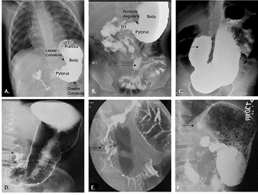
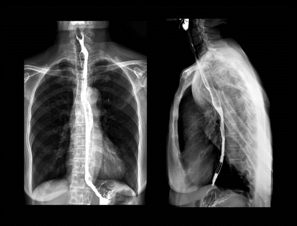
BARIUM SWALLOW
Barium swallow is a dedicated test of the pharynx, esophagus, and proximal stomach, and may be performed as a single or double contrast study. The study is often “modified” to suit the history and symptoms of the individual patient, but it is often useful to evaluate the entire pathway from the lips to the gastric fundus.
BARIUM ENEMA
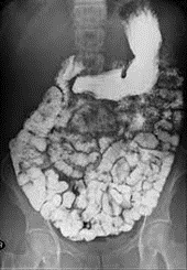
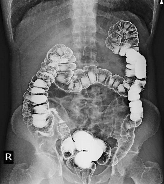
A barium enema is an X-ray exam that can detect changes or abnormalities in the large intestine (colon). The procedure is also called a colon X-ray. An enema is the injection of a liquid into your rectum through a small tube. In this case, the liquid contains a metallic substance (barium) that coats the lining of the colon. Normally, an X-ray produces a poor image of soft tissues, but the barium coating results in a relatively clear silhouette of the colon. During a barium enema exam, air may be pumped into the colon. The air expands the colon and improves the quality of images. This is called an air-contrast (double-contrast) barium enema. Before a barium enema, your doctor will instruct you to completely empty your colon. Call or come for booking for preparation.
HSG
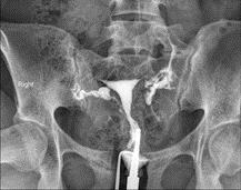
Hysterosalpingography, or HSG, is an X-ray test to outline the internal shape of the uterus and show whether the fallopian tubes are blocked. In HSG, a thin tube is threaded through the vagina and cervix. A substance known as contrast material is injected into the uterus.
One is advised to call or come for booking instructions prior to the procedure.

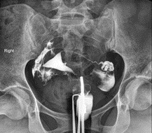
MCUG (micturating cystourethrogram)
A micturating cystourethrogram (MCUG) is a scan/XRAY that shows how well your bladder works by injecting contrast into your bladder through your urethra using a catheter/cannula with images taken as you micturate/urinate. Several images are taken. It is used to diagnose various urinary tract disorders including pain while urinating and difficulty in urinating. It is also used to show up any abnormalities with your urinary system for both adults and children.
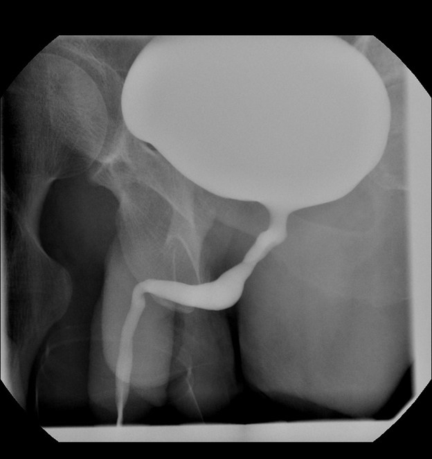
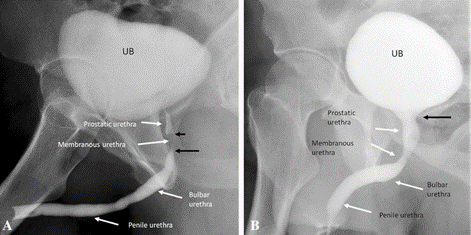
ASCENDING URETHROGRAM
Ascending urethrogram has been the gold standard investigation in the evaluation of stricture urethra. But it is associated with radiation exposure and determines the length and site of the stricture. Done in a retrograde fashion, contrast is injected using a cannula through the urethra into the bladder.
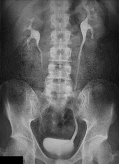
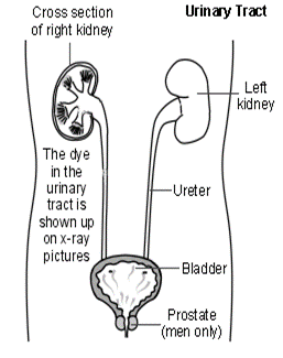
INTRAVENOUS UROGRAPHY
Intravenous urography (IVU), also referred to as intravenous pyelography (IVP) or excretory urography (EU), is a radiographic study of the renal parenchyma, pelvicalyceal system, ureters and the urinary bladder.