Category
Routine Imaging
Synonyms/Aliases
GALACTOGRAPHY, DUCTOGALACTOGRAPHY
Abbreviations
-N/A
Report
1 hour (after completion of the test)
Setup
All working days
Scan
10 MINUTES
Test in Brief
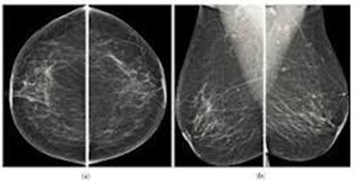
A mammogram is an X-ray picture of the breast. Doctors use a mammogram to look for early signs of breast cancer
Exam Requirements
Try not to have your mammogram the week before you get your period or during your period. Your breasts may be tender or swollen then.
On the day of your mammogram, dont wear deodorant, perfume, or powder. These products can show up as white spots on the X-ray.
Some women prefer to wear a top with a skirt or pants, instead of a dress. You will need to undress from your waist up for the mammogram..
Instructions
You will stand in front of a special X-ray machine. A technologist will place your breast on a clear plastic plate. Another plate will firmly press your breast from above. The plates will flatten the breast, holding it still while the X-ray is being taken. You will feel some pressure. The steps are repeated to make a side view of the breast. The other breast will be X-rayed in the same way. You will then wait while the technologist checks the four X-rays to make sure the pictures do not need to be re-done. Keep in mind that the technologist cannot tell you the results of your mammogram.. Images of good radiographic quality are printed & given to the radiologist for reporting. Used to diagnose abnormalities of the breast (one) e.g. tumours.
Method
You will stand in front of a special X-ray machine. A technologist will place the cannulated breast on a clear plastic plate. Another plate will firmly press your breast from above. The plates will flatten the breast, holding it still while the X-ray is being taken. You will feel some pressure. The steps are repeated to make a side view of the breast. The other breast will be X-rayed in the same way. You will then wait while the technologist checks the four X-rays to make sure the pictures do not need to be re-done. Keep in mind that the technologist cannot tell you the results of your mammogram.. Images of good radiographic quality are printed & given to the radiologist for reporting. Used to diagnose abnormalities of the breast (one) e.g. tumours.
Usefulness /Advantages
Reduces the risk of dying from breast cancer
Of 1,000 women who have a mammogram every 2 years for 20 years, 7 deaths are prevented
Reduces the risk of having to undergo chemotherapy
Screening often allows for the detection of cancers at an early stage of development. Treatment is then possible without chemotherapy.
Allows women to know the health of their breasts
The vast majority of women (nearly 98 %) will not have breast cancer if their mammograms and additional examinations do not reveal cancers.